
Speak To An Expert
Get clear, personalised advice for your situation.
Jot down a few questions to make the most of your conversation.
Introduction to Thrombosis Diagnosis
Thrombosis is a serious medical condition that occurs when a blood clot forms in a blood vessel, obstructing the normal flow of blood. It can lead to severe health issues, including heart attacks, strokes, or pulmonary embolisms. Diagnosing thrombosis timely and accurately is crucial to effectively manage and treat the condition.
Initial Clinical Evaluation
The diagnosis of thrombosis typically begins with an initial clinical evaluation by a healthcare professional, usually a general practitioner (GP) in the UK. The doctor will review the patient's medical history and ask about symptoms such as pain, swelling, redness, or warmth in the affected area. They may also inquire about risk factors like recent surgery, immobility, or history of clotting disorders.
Physical Examination
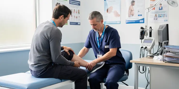
A physical examination is conducted to observe any signs that might indicate thrombosis. The GP will look for swelling, tenderness, or changes in skin colour in the limbs, which are common signs of deep vein thrombosis (DVT). If symptoms suggest thrombosis in the lungs, such as shortness of breath, additional evaluations may be conducted.
Blood Tests
To aid in the diagnosis, the GP might order blood tests, such as a D-dimer test. D-dimer is a protein fragment present in the blood after a blood clot dissolves in the body. Elevated levels of D-dimer might indicate an increased likelihood of thrombotic activity, prompting further investigation. However, a positive D-dimer result is not conclusive by itself and must be paired with other diagnostic techniques.
Imaging Studies
Imaging studies play a vital role in confirming the presence of thrombosis. The most common imaging test for diagnosing DVT is an ultrasound scan, which uses sound waves to visualize the blood flow in the veins and identify any clots. For suspected pulmonary embolism, a CT pulmonary angiography is often performed to get detailed images of the blood vessels in the lungs.
Venography and Other Techniques
In certain cases, a venography may be conducted. This invasive test involves injecting a contrast dye into a vein and then taking X-ray images to detect blockages. MRIs or other advanced imaging techniques may also be used when other methods are inconclusive or when the thrombosis is in an unusual location.
Conclusion
Diagnosing thrombosis involves a combination of clinical evaluations, blood tests, and imaging techniques to ensure accuracy and prompt treatment. Early diagnosis is essential to prevent complications and improve health outcomes. If you suspect you have symptoms of thrombosis, it is crucial to seek medical advice immediately.
What is a Blood Clot?
A blood clot is when blood turns into a lump inside a blood vessel. This lump can block normal blood flow. Blood clots can be very dangerous. They might cause big problems like heart attacks or strokes. It is important to find out if someone has a blood clot quickly so doctors can help them.
Meeting the Doctor
To check for a blood clot, you first meet with a doctor. In the UK, this is often your local doctor or GP. The doctor will ask about your health and look for signs like pain, swelling, or redness in your arms or legs. They will also ask if you have had any recent operations or if you have a history of blood problems.
Body Check
The doctor will look at your body to see if there are signs of a blood clot. They look for swelling or changes in your skin. If you have trouble breathing, they might do more tests to see if there is a blood clot in your lungs.
Blood Tests
The doctor might ask for a blood test called a D-dimer. This test checks for a certain protein in your blood. If there is more of this protein, it might mean you have a blood clot. But, the blood test by itself is not enough to say for sure.
Pictures of Inside the Body
To be sure about a blood clot, doctors use special pictures of your body. The most common test is an ultrasound. It uses sound waves to see the blood flow and find clots in your veins. If the doctor thinks there is a clot in your lungs, they might do a special scan called a CT scan.
Other Tests
Rarely, doctors use a test called venography. This test uses a dye to see your veins with X-rays. Sometimes they use an MRI to look inside the body, especially if the clot is in a hard-to-see place.
Why It Matters
Finding a blood clot early is very important. It helps keep you safe and healthy. If you think you might have a blood clot, tell a doctor right away. They will help you get the right tests and treatment.
Frequently Asked Questions
Common methods include ultrasound, blood tests (such as D-dimer), venography, CT scans, and MRI.
Yes, ultrasound is commonly used to diagnose deep vein thrombosis (DVT) as it can visualize blood flow and blockages in veins.
A D-dimer test measures blood levels of D-dimer, a substance released when a blood clot dissolves, and is used to help diagnose or rule out thrombosis.
Venography involves injecting a contrast dye into a vein, usually in the foot, and taking X-rays to visualize blood flow and detect clots.
CT and MRI scans provide detailed images of blood vessels and can detect blood clots, especially in more complex cases or areas difficult to assess with ultrasound.
Yes, risk factors include prolonged immobility, recent surgery, cancer, pregnancy, and a history of blood clots.
No, symptoms alone are not sufficient for diagnosis; imaging tests or blood tests are necessary to confirm thrombosis.
A positive D-dimer test indicates the need for further testing, as it suggests clot presence but can be elevated due to other conditions.
If initial tests are inconclusive or risk factors are high, further testing provides more detail to confirm the presence of a clot.
Yes, thrombosis can be asymptomatic, which is why diagnostic tests are crucial if there is high suspicion.
Yes, a thorough patient history can identify risk factors and guide the decision-making process for testing.
Ultrasound is highly reliable for detecting clots in the legs or arms, particularly for DVT, with a high sensitivity and specificity.
Typically, no special preparations are needed for ultrasounds, but fasting may be required for some contrast-enhanced scans like CT.
Most tests are non-invasive and safe, but venography involves exposure to radiation and contrast dye, which carries minor risks.
Results can be immediate for ultrasounds, while blood tests and advanced scans may take several hours to process and review.
Yes, factors such as inactivity, diet, and habits like smoking can affect clot formation and risk perception, thus influencing diagnostic testing.
Physical examination can help identify symptoms indicative of thrombosis, such as swelling or pain, guiding further diagnostic testing.
Untreated thrombosis can lead to serious complications, such as pulmonary embolism, which may be life-threatening.
Yes, due to physiological changes, pregnant women may require tailored testing protocols to avoid undue risks from radiation exposure.
Patients with recurrent thrombosis may undergo more frequent monitoring and extensive testing strategies to manage and prevent further episodes.
Doctors use different ways to look inside your body. They might use:
- A special camera called an ultrasound.
- A blood test. One kind is the D-dimer test.
- Pictures of your veins, called venography.
- A CT scan, which is like a special X-ray.
- An MRI, which takes pictures inside your body.
You can use pictures, videos, or ask someone to explain these if it's hard to understand.
Yes, doctors use a special tool called an ultrasound to look inside the body. It helps them see blood flow and find clots in veins. This is called deep vein thrombosis or DVT.
A D-dimer test checks your blood for D-dimer. D-dimer is something that comes out when a blood clot goes away. Doctors use this test to find out if you have a problem with blood clots.
Venography is a test to look at blood flow. The doctor puts a special dye in your foot. Then, they take pictures to see if there are any blood clots.
CT and MRI scans take pictures of blood vessels. They can find blood clots, even in tricky places where an ultrasound might not work well.
Yes, there are things that can make it more likely for you to get blood clots. These include sitting still for a long time, having surgery recently, having cancer, being pregnant, and if you have had blood clots before.
No, just feeling sick is not enough to know if you have a blood clot. Doctors need to do special tests, like taking pictures of inside your body or checking your blood, to be sure.
A positive D-dimer test means we might need to do more tests. This is because it can show there might be a blood clot. But sometimes it can be high for other reasons too.
If the first tests do not give a clear answer or if there is a high risk, more tests can help find out if there is a clot.
Yes, thrombosis can happen without any symptoms. This is why it is important to have tests if doctors think you might have it.
Yes, asking the patient lots of questions can help find out what might be a problem. It can also help doctors decide if any tests are needed.
Ultrasound is very good at finding blood clots in the legs or arms. It is especially good at finding deep vein thrombosis (DVT). It is almost always right when it says there is a clot.
Usually, you don't need to do anything special to get ready for an ultrasound. But, for some scans called CT scans, you might need to not eat or drink before. This is called fasting.
Most tests are safe and don't go inside your body. But venography is a test that uses a special camera and a dye. It is a little more risky because of the camera's rays and the dye.
You can get results right away with an ultrasound. But with blood tests and special scans, it might take a few hours to get the answers.
Yes, things like not moving around much, what you eat, and habits like smoking can change how blood clots form. This also affects how doctors check for these clots.
Doctors check your body to find signs of a blood clot, like if it's swollen or hurts. This helps them know if more tests are needed.
If thrombosis is not treated, it can cause big problems. One problem is a pulmonary embolism, which can be very dangerous and might even be life-threatening.
Yes, when women are having a baby, their bodies change. They might need special tests to keep them safe from too much radiation.
People who get blood clots often might need to see the doctor more and have extra tests to help stop more clots from happening.

Ergsy Search Results
This website offers general information and is not a substitute for professional advice.
Always seek guidance from qualified professionals.
If you have any medical concerns or need urgent help, contact a healthcare professional or emergency services immediately.
Some of this content was generated with AI assistance. We've done our best to keep it accurate, helpful, and human-friendly.
- Ergsy carefully checks the information in the videos we provide here.
- Videos shown by Youtube after a video has completed, have NOT been reviewed by ERGSY.
- To view, click the arrow in centre of video.
- Most of the videos you find here will have subtitles and/or closed captions available.
- You may need to turn these on, and choose your preferred language.
- Go to the video you'd like to watch.
- If closed captions (CC) are available, settings will be visible on the bottom right of the video player.
- To turn on Captions, click settings.
- To turn off Captions, click settings again.