
Speak To An Expert
Get clear, personalised advice for your situation.
Jot down a few questions to make the most of your conversation.
Can ultrasound see through bone?
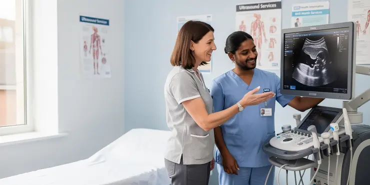
Ultrasound is very good at creating images of soft tissues, such as muscles, tendons, ligaments and some organs. However, it cannot properly see through bone in the same way that it can pass through softer body tissues. Bone blocks and reflects most of the sound waves, so the image behind it is usually unclear.
This means a standard ultrasound scan is not the right tool for looking inside solid bones. If a clinician needs to check bone structure, they will usually consider an X-ray, CT scan or MRI instead, depending on the question they need to answer.
Why bone is a problem for ultrasound
Ultrasound works by sending high-frequency sound waves into the body and measuring the echoes that bounce back. Soft tissues let many of those waves travel through, which allows the machine to build up an image. Bone is much denser, so it reflects most of the waves at the surface.
Because of this, the ultrasound probe can only show the outer edge of the bone clearly. Anything deep to the bone is often hidden, or appears as a shadow on the screen. That is why ultrasound is limited when it comes to examining fractures or conditions inside the bone itself.
What ultrasound can still show around bone
Although ultrasound cannot see through bone, it can still be very useful near bones. It can show muscles, tendons, joints and fluid collections around the bone. It is commonly used to help assess injuries, inflammation and some soft tissue problems.
In some cases, ultrasound can also help guide procedures, such as injections or fluid drainage, especially around joints. In the UK, it is often used because it is quick, painless and does not involve ionising radiation. This makes it a practical option for many routine checks.
When other scans are better
If a doctor suspects a broken bone, an X-ray is usually the first scan requested. X-rays are much better for showing bone detail and are widely available in NHS settings. If more detail is needed, CT scans can give a more complete picture of complex fractures.
MRI scans are useful when the concern is not just the bone, but also the marrow, cartilage or surrounding soft tissues. The best scan depends on the part of the body being examined and the condition being investigated. A clinician will choose the most suitable test for the situation.
The bottom line
Ultrasound cannot truly see through bone. It can only show the surface of the bone and the soft tissues around it.
That does not make it less useful. It simply means it is best used for different tasks, while X-rays, CT and MRI are better for looking inside or beyond bone.
Frequently Asked Questions
Ultrasound see through bone refers to the use of high-frequency sound waves to visualize structures beneath or around bone, often by looking through softer tissue windows or by using advanced processing. Because bone strongly reflects and absorbs ultrasound, it generally cannot be truly "seen through" in a literal sense, but useful images can sometimes be obtained around thin bone, at bone edges, or through acoustic windows.
Ultrasound see through bone in adults is usually very limited because adult bone blocks most ultrasound waves. In many cases, clinicians instead image around bone or use ultrasound for nearby soft tissues, joints, vessels, or guided procedures where a soft-tissue path exists.
Ultrasound see through bone in infants and children can be more feasible than in adults because their bones are thinner and some skull sutures and fontanelles remain open. This is why ultrasound is sometimes used for neonatal brain imaging and other pediatric applications.
Ultrasound see through bone poorly because bone has a very different acoustic impedance than soft tissue. That mismatch causes most of the sound wave to reflect, scatter, or be absorbed, leaving little energy to form an image beyond the bone.
Ultrasound see through bone works best where bone is thin, incomplete, or separated by openings such as the infant skull fontanelle, temporal acoustic windows, or thin cortical areas. It is also more practical near joints and edges of bone where sound can reach the target through adjacent soft tissue.
Ultrasound see through bone to image the brain is generally difficult in adults because the skull blocks the beam. In newborns and some special circumstances, such as through a fontanelle or an acoustic window, ultrasound can provide useful brain images.
Ultrasound see through bone to detect fractures cannot reliably image the interior of bone, but it can sometimes identify surface irregularities, cortical breaks, or associated soft-tissue swelling. It is not a complete replacement for X-ray, CT, or MRI when a fracture is suspected.
Ultrasound see through bone for joint problems can be useful because many joints have accessible soft-tissue margins. It can show joint effusions, tendon injuries, bursitis, and some cartilage-related findings, although the deeper parts of a joint may still be obscured by bone.
Ultrasound see through bone during surgery or procedures is often used to guide needles, drains, and nerve blocks near bony landmarks rather than through solid bone itself. It helps clinicians avoid surrounding structures and improve accuracy when a soft-tissue path is available.
The main limitations of ultrasound see through bone are strong reflection from bone, shadowing behind the bone, limited penetration, and reduced image quality. Air, thick cortical bone, and deep targets further reduce usefulness.
Ultrasound see through bone is generally considered safe when used appropriately because it does not use ionizing radiation. As with any ultrasound exam, it should be performed by trained professionals who follow standard safety and clinical guidelines.
Ultrasound see through bone does not usually outperform X-ray or CT for looking at bone itself. X-ray and CT are much better for visualizing bone structure, while ultrasound is better for soft tissues and certain guided procedures near bone.
Ultrasound see through bone can sometimes help detect soft-tissue abscesses, fluid collections, or inflammation near bone, but it cannot reliably assess infection inside bone. For suspected osteomyelitis or deep bone infection, MRI, CT, or other tests are often needed.
Ultrasound see through bone can sometimes measure blood flow in vessels near bones if the vessel is accessible from a soft-tissue angle. Doppler ultrasound is commonly used for this purpose, though thick bone may block the view.
An acoustic window in ultrasound see through bone is an area where sound waves can pass with less interference, such as a thin bone region or natural opening. These windows allow clinicians to image deeper structures that would otherwise be hidden by bone.
Ultrasound see through bone in the skull is limited because the skull is dense and highly reflective. It can be possible through thinner areas, open fontanelles in infants, or specific skull windows, but not through most of the adult skull.
Ultrasound see through bone is less capable than MRI for viewing structures beyond bone. MRI can image many deep tissues without being blocked by bone, while ultrasound is fast, portable, and useful when the target is accessible from a soft-tissue path.
Ultrasound see through bone is useful in emergency medicine for evaluating fluids, soft-tissue injuries, pneumothorax, vascular access, and some pediatric head scans. It is especially valuable when quick bedside imaging is needed and the target is reachable around bone.
Ultrasound see through bone can be improved somewhat with advanced transducers, image processing, and specialized reconstruction methods, but it still faces fundamental physical limits. These technologies may enhance images near or around bone, but they do not make solid bone transparent to ultrasound.
During an ultrasound see through bone exam, a technician places gel and a probe on the skin and moves it to find a usable acoustic window. Patients usually feel only mild pressure, and the exam is noninvasive, but the provider may explain that bone may limit image quality.

Ergsy Search Results
This website offers general information and is not a substitute for professional advice.
Always seek guidance from qualified professionals.
If you have any medical concerns or need urgent help, contact a healthcare professional or emergency services immediately.
Some of this content was generated with AI assistance. We've done our best to keep it accurate, helpful, and human-friendly.
- Ergsy carefully checks the information in the videos we provide here.
- Videos shown by Youtube after a video has completed, have NOT been reviewed by ERGSY.
- To view, click the arrow in centre of video.
- Most of the videos you find here will have subtitles and/or closed captions available.
- You may need to turn these on, and choose your preferred language.
- Go to the video you'd like to watch.
- If closed captions (CC) are available, settings will be visible on the bottom right of the video player.
- To turn on Captions, click settings.
- To turn off Captions, click settings again.